September 7, 2024
Ultrasound Therapy: What Is It, How It Works, Benefits
What To Expect During A Restorative Ultrasound Analysis ultrasound is often identified by the facility regularity of the pulses (commonly in the 2-- 12 MHz array), which is typically a regularity integral to the density of the ceramic crystal. As the pressure amplitude, the regularity, or the proliferation size is enhanced, the ultrasound wave can distort, which might inevitably cause an interruption or shock in the waveform. In relation to bioeffects, raising regularity, nonlinear acoustic distortion, or pulse length can enhance home heating and enhance some nonthermal mechanisms, e.g., radiation pressure. Decreasing regularity enhances the possibility of cavitation and gas body activation. Increasing power or strength tends to enhance the possibility and magnitude of all bioeffects mechanisms. Restorative ultrasound devices might make use of brief bursts or constant waves to deliver reliable ultrasonic power to cells.- Depending upon the condition, ultrasound (energetic and placebo) was used alone or along with various other interventions in a way developed to determine its contribution and differentiate it from those of various other interventions.
- Besides my teaching at the college and being a researcher, I am likewise a facility owner.
- One of the most commonly studied bone and joint concerns is nerve entrapment with carpal tunnel syndrome (CTS).
- Lithotripsy triggers injury to basically all patients (Evan and McAteer, 1996).
- A professional system has actually been authorized for fat debulking in the European Union and Canada (Fatemi, 2009).
Exactly how commonly can you utilize ultrasound therapy?
Just how commonly can you make use of ultrasound treatment? Ultrasound treatment can be used as commonly as essential, there are no restrictions. We normally utilize it for 5 minutes each time during treatment. Whether we use it or otherwise will certainly depend upon the client''s injuries.
What Are The Dangers Of Restorative Ultrasound?
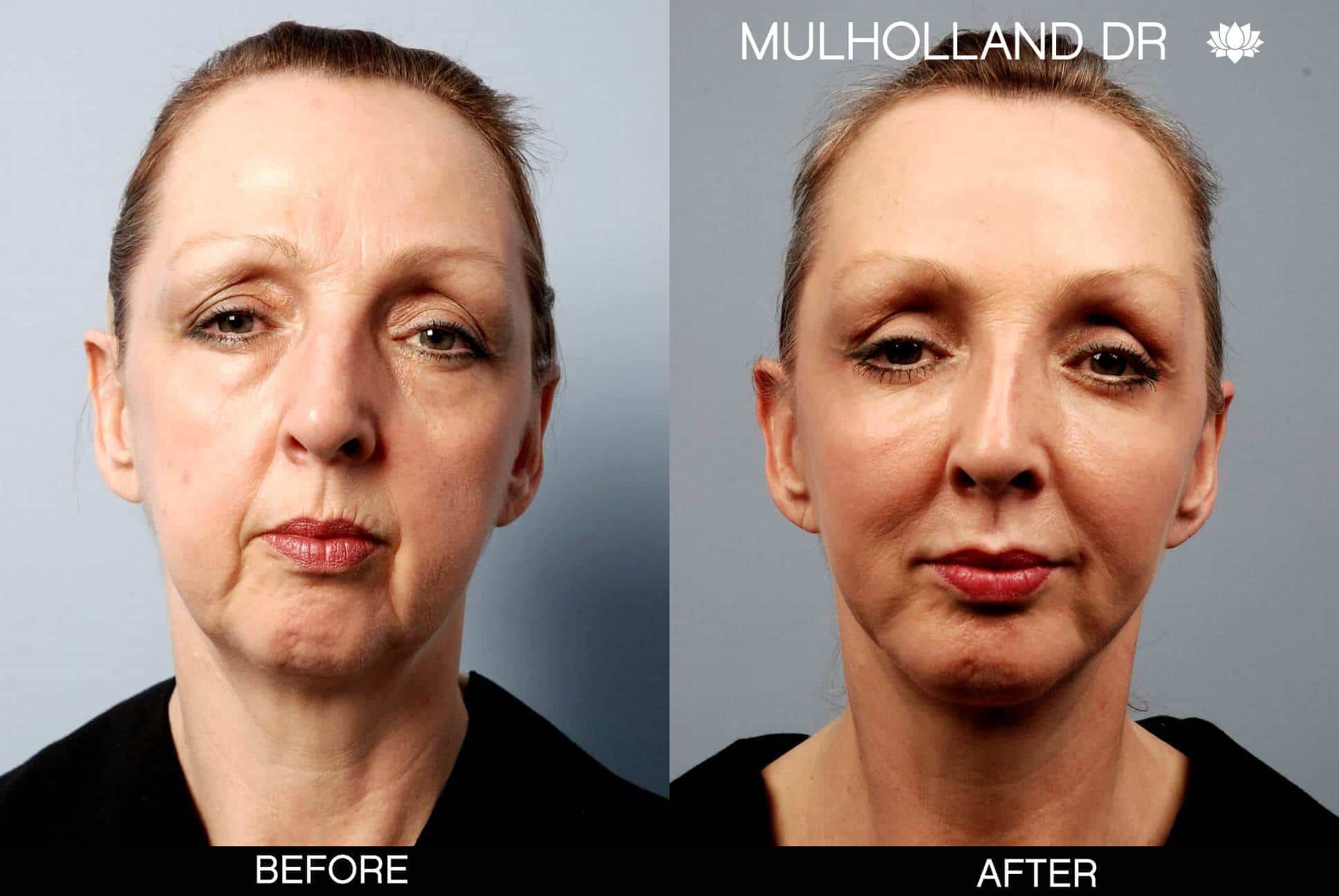
Really little sores of ~ 1 mm3 up to numerous 10s of cm3 can be produced. The method might supply a much safer choice to liposuction surgery for aesthetic applications (Moreno-Morega et al. 2007). Shallow cells is revealed to HIFU leading either to a tightening of collagen centered cells (dermis) or to devastation of fat (Gliklich et al. 2007; White et al. 2007).Advantages Of
The duration of this discomfort normally fixes within a couple of hours, but it can occasionally persist for approximately 24 to two days. It's your body's way of reacting to the therapy, and it typically indicates that the recovery process has begun. Ultrasound makes it possible for doctor to "see" information of soft cells inside your body without making any type of incisions (cuts). We included randomised controlled tests (RCTs) on healing ultrasound for chronic non-specific LBP. We compared ultrasound (either alone or in mix with one more therapy) with sugar pill or other treatments for chronic LBP. Diagnostic ultrasounds utilize acoustic waves to make photos of the body. A professional system has been approved for fat debulking in the European Union and Canada (Fatemi, 2009). Depending on the gadget, as well as the cosmetic application, both thermal along with non-thermal systems within an ultrasound area are used for these procedures. One of these tools is currently accepted for scientific usage in the USA (Alam et al. 2010), and others are in usage worldwide. Long term application of this innovation, as well as governing approval, is still progressing. A radiologist can measure the size and color versus various other close-by organs to contrast them. Projections or spots on the surface might suggest cysts or strong masses. A radiologist could additionally try to find bigger (dilated) blood vessels or bile ducts. Your sonographer will certainly send the images from your examination to a radiologist.Social Links